


Radiology
The Radiology Department provides you with services, equipped with all the necessary expertise and technology.
Expertise in Imaging Technologies:
Radiology Department
Dear Patient and their Relatives;
Our hospital, established in 1898 and built on a rich legacy, takes great pride in providing healthcare services to the community for over a century. Our Radiology Department is here to support the diagnosis and treatment processes of our patients, with our expert team and advanced imaging technologies.
Our Radiology Department offers the latest medical imaging technologies to our patients. We use devices such as MRI (Magnetic Resonance Imaging), CT (Computed Tomography), X-ray, ultrasound, and mammography to diagnose various conditions. Our expert radiologists carefully analyse the obtained images to ensure the accuracy of the diagnoses and to develop appropriate treatment plans.
-
 Radiology Services

Our Doctors
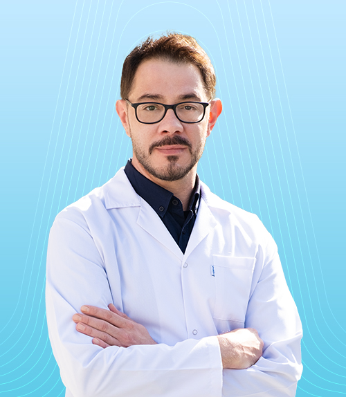
M.D FIRAT KESKİNER, Specialist
RADIOLOGY
Get Detailed Information About the Radiology Department
It has been prepared by the doctors of our hospital.
Interventional radiology is a medical field that uses imaging techniques (such as ultrasound, X-rays, computed tomography, or magnetic resonance imaging) to perform therapeutic procedures within the body. This approach helps treat diseases in a less invasive way, often eliminating the need for surgery.
How Is It Done?
Interventional radiologists use imaging technology to precisely locate the affected area and perform treatment with thin needles or tubes. These procedures are typically carried out under local anaesthesia, meaning the patient remains awake and only the treatment area is numbed.
Applications of Interventional Radiology
- Vascular Blockages and Stent Placement:
- Stents are placed to open blocked vessels and restore proper blood flow.
- Tumour Treatments:
- Drugs may be delivered via needle, or thermal ablation can be performed to destroy cancerous cells.
- Liver, Kidney, and Other Organ Biopsies:
- Suspicious masses can be sampled for diagnosis.
- Clearing Obstructed Bile Ducts:
- Blocked bile ducts can be opened to restore bile flow.
- Bleeding Control:
- Internal bleeding can be stopped by treating the blood vessels involved.
- External Catheter Placement:
- Catheters may be placed for conditions such as kidney stones.
Advantages
- Faster recovery times
- Lower risk due to the absence of major surgical incisions
- Typically, shorter hospital stays
- Less pain for the patient
Important Note:
Interventional radiology can serve as an excellent alternative to surgery. For any medical procedures, it’s crucial to seek treatment recommendations from a specialized radiologist.
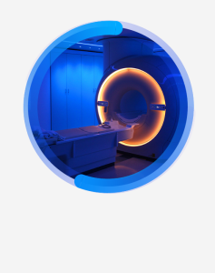
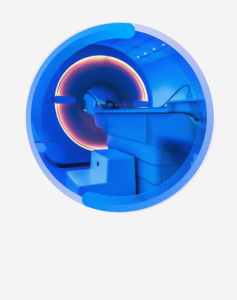
Magnetic resonance imaging (MRI) is a diagnostic method used to visualize structures inside the body. By employing magnetic fields and radio waves, MRI generates highly detailed images of internal tissues. It is particularly valuable for examining soft tissues such as the brain, spinal cord, muscles, heart, and various organs.
How Does It Work?
The MRI machine uses a strong magnetic field to affect hydrogen atoms in the body. These atoms respond to radio waves and emit signals that are processed into detailed images displayed on a computer screen. As a result, internal structures become clearly visible.
Applications of MRI
- Brain and Spinal Cord:
- Used to diagnose conditions like brain tumours, vascular issues, and multiple sclerosis (MS).
- Joints and Muscles:
- Evaluates injuries in joints like the knee or shoulder, as well as muscle problems and ligament tears.
- Heart and Blood Vessels:
- Monitors heart diseases, vascular blockages, and blood flow.
- Liver and Other Organs:
- Assesses diseases or lesions in internal organs such as the liver, kidneys, and pancreas.
Advantages
- High Resolution: Provides highly detailed imaging of soft tissues.
- Radiation-Free: Unlike X-rays, MRI does not use harmful radiation.
- Non-Invasive: MRI does not involve inserting any devices into the body.
Important Note:
MRI is a highly useful tool for diagnosing many neurological, cardiac, and orthopaedic conditions. If an MRI is deemed necessary, your doctor will plan the most appropriate test for you.
Computed tomography (CT) is an imaging technique used to obtain detailed images of internal structures within the body. By utilizing X-rays, CT produces cross-sectional views of the body, which are then processed by a computer to create three-dimensional images.
How Does It Work?
A CT scanner emits X-rays into the body and collects the rays reflected back from different tissues. This data is then processed by a computer to produce multiple cross-sectional images. As a result, internal organs, bones, and other structures can be examined in detail.
Applications of CT
- Trauma and Post-Accident Conditions:
- Used to evaluate bone fractures, internal organ injuries, or head trauma.
- Brain:
- Can detect brain haemorrhages, tumours, and stroke.
- Lungs:
- Evaluates lung diseases, cancers, and infections.
- Internal Organs:
- Allows for the identification of diseases or masses in organs such as the liver, kidneys, and pancreas.
- Cancer Screening:
- Used to determine the location, spread, and size of cancers.
Advantages
- Fast Results: CT can produce images quickly, saving time in emergencies.
- High Resolution: Provides high-quality images of bones and internal organs.
- Detailed Visualization: Offers three-dimensional cross-sections, enabling more thorough examinations.
Important Note:
While CT is very effective for rapid diagnosis in certain conditions, it does involve the use of X-rays and should be used judiciously. If a CT scan is recommended, your doctor will provide the necessary guidance and information.
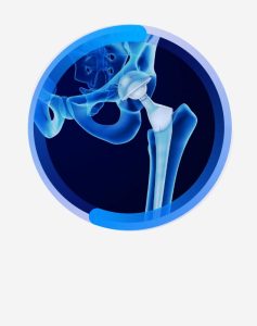
X-rays are a commonly used medical test that allows doctors to visualize internal structures within the body. By directing X-ray beams into the body, a picture is created based on how different tissues absorb and reflect the radiation. X-rays are particularly useful for quickly assessing bones and certain organs.
How Does It Work?
An X-ray machine emits low-dose radiation beams through the body. These beams are absorbed at different rates depending on the density of the tissues. As a result, bones appear white, while softer tissues show up in shades of grey. The resulting image helps identify the location and condition of diseases or injuries.
Applications of X-Ray Imaging
- Bone Fractures and Dislocations:
- Quickly evaluates the condition of bones after accidents or trauma.
- Lung Conditions:
- Used to assess lung infections, tuberculosis, pneumonia, and similar diseases.
- Dental Health:
- Detects cavities, root issues, and other dental problems.
- Abdomen and Internal Organs:
- Provides information about abdominal organs, such as the stomach and intestines.
- Cancer Screening:
- May be used for early detection of certain types of cancer.
Advantages
- Quick and Convenient: X-ray procedures are fast, often providing same-day results without a hospital visit.
- Cost-Effective: X-rays are generally more affordable than other imaging methods.
- Versatile: Can quickly identify changes in bones and certain organs.
Important Note:
X-rays typically use low doses of radiation, but it is important to avoid unnecessary exposure. If an X-ray is needed, your doctor will provide you with all necessary information.
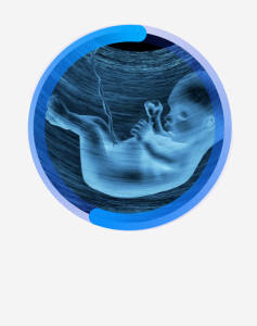
Ultrasound (USG) is a test that uses sound waves to produce images of the body’s internal structures. It is often employed to assess organs, tissues, and blood vessels. Colour Doppler ultrasound, a specialized type of ultrasound, examines the movement of blood within vessels. This method provides color-coded images that show blood flow and the condition of blood vessels.
Applications of Ultrasound
- Pregnancy Monitoring: Observing the baby’s growth, position, and health.
- Abdomen and Internal Organs: Examining organs such as the liver, kidneys, and gallbladder.
- Musculoskeletal System: Assessing joints, muscles, tendons, and ligaments.
- Thyroid and Lymph Nodes: Checking for suspicious masses.
- Heart and Blood Vessels: Analysing the structure of the heart and identifying certain vascular conditions.
Applications of Colour Doppler Ultrasound
- Blood Flow Assessment: Visualizing the speed and direction of blood flow within vessels.
- Vascular Obstructions and Clots: Determining how smoothly blood is moving through vessels.
- Varicose Veins: Evaluating the condition of large veins in the legs.
- Cardiac Vessels: Identifying blockages and flow disturbances in the heart’s vessels.
- Carotid Arteries: Assessing blockages or narrowing in the arteries supplying blood to the brain.
Advantages
- Non-Invasive: Produces internal images without the need for any surgical intervention.
- Safe and Fast: Unlike X-rays, it does not involve radiation and is considered safe.
- Real-Time Imaging: Allows immediate visualization of blood flow inside vessels.
- Cost-Effective: Generally, more affordable compared to other imaging methods.
Important Note:
Ultrasound and Colour Doppler ultrasound play a crucial role in both diagnosis and treatment. They are usually low-risk and provide quick results. If you need these tests, your doctor will select the most appropriate one and guide you accordingly.
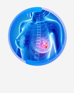
Mammography is the process of imaging breast tissue using X-rays. It is commonly used for the early detection of breast cancer, as well as for identifying lumps, cysts, and other abnormal conditions in the breast. Mammography is a standard routine screening tool, particularly for women.
Applications of Mammography
- Breast Cancer Screening: Used to detect cancer in its early stages.
- Examination of Breast Masses: Determines whether a suspicious lump is benign or malignant.
- Monitoring in High-Risk Individuals: Recommended for individuals with a family history of breast cancer to undergo regular mammograms.
- Evaluation of Breast Changes: Assesses symptoms such as pain, swelling, or growth in breast tissue.
Advantages of Mammography
- Early Detection: Helps identify breast cancer at an early stage, increasing treatment success.
- Quick and Effective: Provides accurate results in a short amount of time.
- Non-Invasive: A painless, non-surgical procedure.
Mammography Screening Recommendations
- Women aged 40 and above are advised to have a mammogram annually or every two years.
- Those with a family history of breast cancer may need mammograms at a younger age.
- If there are any abnormal symptoms (lumps, pain, discharge, etc.), mammography can be performed as recommended by a doctor.
Important Note:
Mammography is crucial for the early detection of breast cancer. It should be performed at regular intervals within a specified age range. Mammogram results should be reviewed by a physician, and further tests may be recommended if necessary.
Our hospital provides 24-hour uninterrupted service with a team consisting of 1 Radiology Doctor, 6 Radiology Technicians, 1 Secretary, and 1 Support Staff.
At our hospital, the following imaging services are available:
- MRI (Magnetic Resonance Imaging) 1.5 Tesla Siemens
- CT (Computed Tomography) 16-slice, 3D imaging Siemens
- X-ray
- Mammography
- Ultrasound
- Various imaging procedures are performed using these advanced technologies.
Health Library

